History: 14 year old female with right lower quadrant pain

Axial LAVA-Flex MRI demonstrates a pelvic cystic structure containing fat (white arrow) as indicated by the signal corresponding to that of the subcutaneous fat
Axial FIESTA demonstrates a pelvic cystic structure containing fat (white arrow) as indicated by the signal corresponding to that of the subcutaneous fat
Axial T2W MRI demonstrates a pelvic cystic structure containing fat (white arrow) as indicated by the signal corresponding to that of the subcutaneous fat

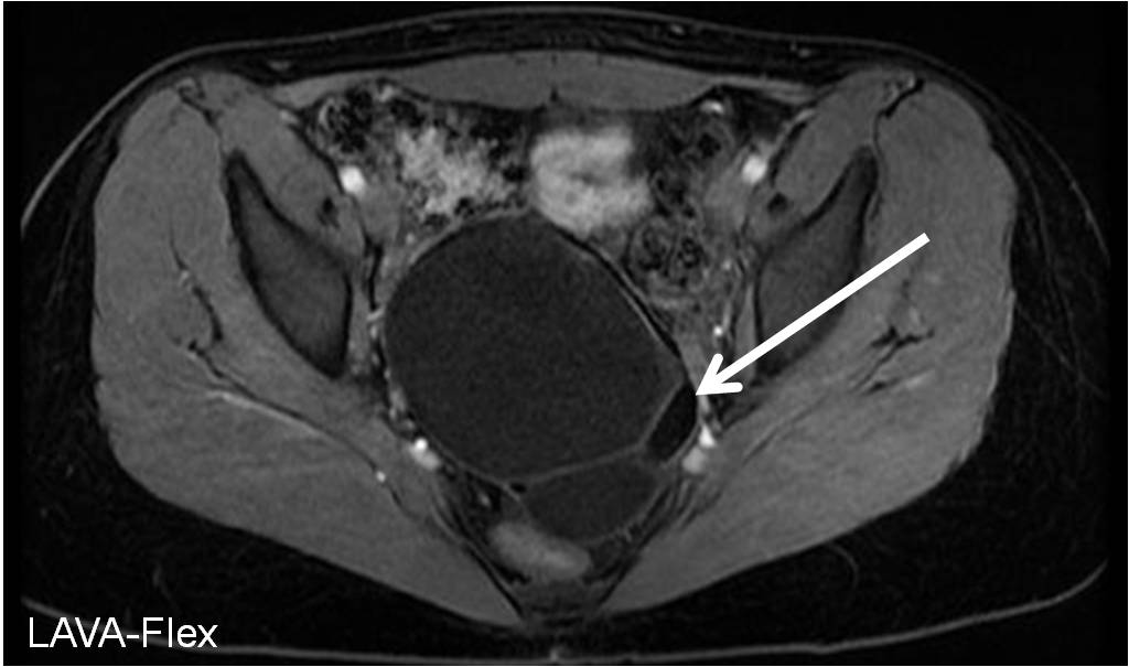
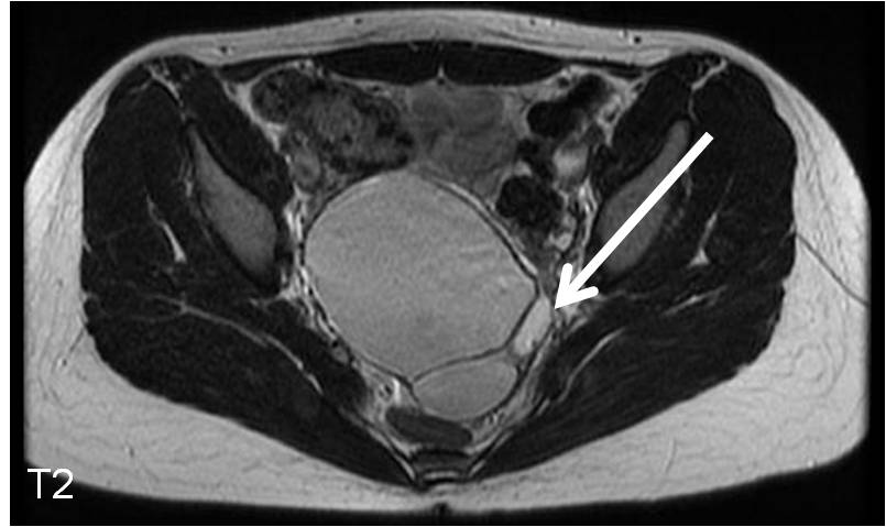
This is the appearance of an ovarian dermoid cyst on MRI, also known as a mature teratoma. The name teratoma gives a clue as to the histology of the dermoid cyst, usually containing parenchymal cell types from all three germ cell layers (mesoderm, endoderm, and ectoderm). The ectoderm components are those of typical skin structures, including hair and fat (as seen in the images above). Dermoid cysts may also contain teeth.

See an ovarian fibroma on ultrasound.

Leave a comment

Trending