

A) Dermoid plug CORRECT
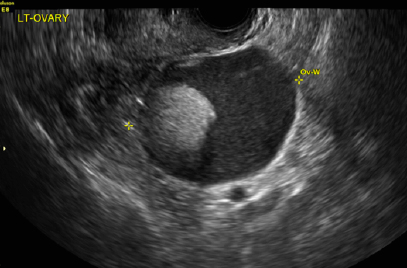
The most common appearance of an ovarian dermoid is a cystic lesion with a focal echogenic nodule protruding into the cyst (Rokitansky nodule).1
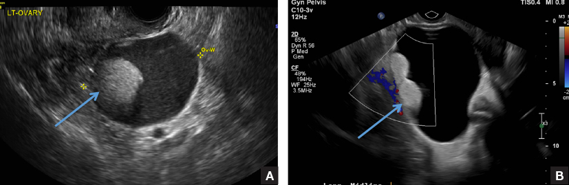
Transvaginal pelvic ultrasounds on 2 different patients demonstrate focal echogenic nodules (long arrows) protruding into the cyst (Rokitansky nodule).
B) Tip-of-the-iceberg sign INCORRECT
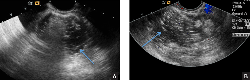
The next most common appearance of an ovarian dermoid is a focal or diffuse hyperechoic mass with areas of sound attenuation from the sebaceous material and hair, often called the tip-of-the-iceberg sign.1
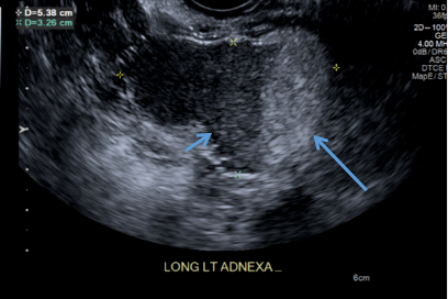
Transvaginal pelvic ultrasounds from 2 different patients demonstrate focal hyperechoic masses (long arrows) with areas of sound attenuation (arrowheads) precluding delineation of the entire dermoid.
C) Dot-dash pattern INCORRECT
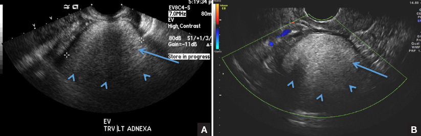
The 3rd most common appearance of an ovarian dermoid is a cystic lesion with multiple thin echogenic bands (lines and dots) that visualize hair floating within the cyst.1
Transvaginal pelvic ultrasounds of the right ovary (transverse and longitudinal views of the same ovary) demonstrate a cystic lesion with multiple thin echogenic bands (lines and dots) showing hair floating within the cyst (long arrows).
D) Fat-fluid level INCORRECT
The 4th most common appearance of an ovarian dermoid is a result of the echogenic sebum and hypoechoic serous fluid causing a fat-fluid level.1
Transvaginal pelvic ultrasound of the left ovary demonstrates a cystic lesion with echogenic sebum (long arrow) and hypoechoic serous fluid causing a fat-fluid level (short arrow).